
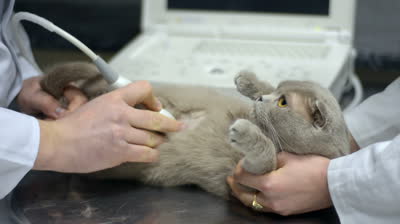
In the month of December, we would like to bring awareness to the importance of basic full abdominal ultrasound scans!
An ultrasound scan of the full abdomen usually takes between 15-45min depending on individuals and the usual price is $250.
- Please note that this does not include consultation fees for new patients, blood tests, surgery procedure itself, x-rays or any other tests that may be required. It is highly preferred that x-rays are taken prior to an ultrasound.
- All ultrasound patients will need to have the belly shaved in order to get good quality images. This will be done in the clinic as a complimentary service. Only the area of interest will be shaved.
- No ultrasound reports will be provided during the month of December but owners can get a copy of the labelled images via email.
- We apologize that due to time constraints, owners will not be allowed to watch the ultrasound performed during this month. We will try out best to send the images and videos of your pet's scan via email within 14 working days.
- We retain the right to refuse performing a scan on patients that are not stable during surgery/anaesthesia or patients who are considered high risk (heart problem, liver failure etc). The patients' well being and health remains the top priority.
What is the difference between x-rays and ultrasound?
Radiographs, or x-rays, are an essential first step in order to get the "big picture" of what is going on inside your pet's body. It can be used to diagnose an enlarged heart, assess liver size and margins, check for tumours and growths within the body and evaluate dental disease and bone health. However, xrays are only able to provide a 2D picture of what is happening.
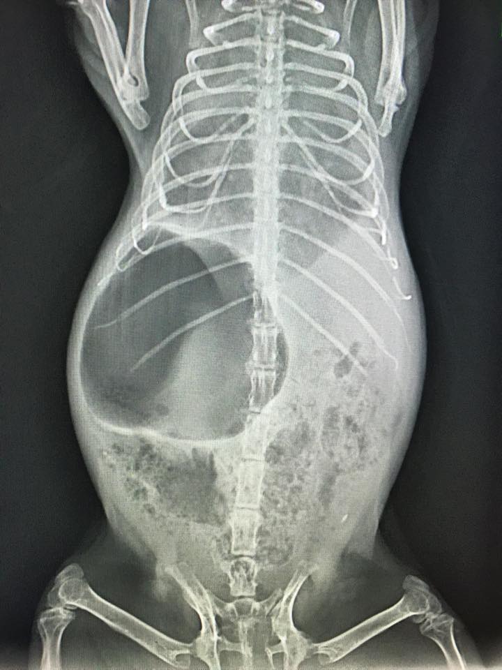
Guinea pig with severe pneumonia (lungs would normally show up as black when they are filled with air). She was also suffering from gut stasis and gastric dilation (a lot of black gas seen in stomach).
Ultrasound imaging, on the other hand, allows us to obtain more in depth details. Organ texture and certain organs and glands that would not normally be seen on x-rays can also be assessed with ultrasonography. These include blood flow supply to the kidneys to assess kidney health, lymph node size, adrenal glands for hormonal problems and bladder stones and sand.

Gall bladder with a mucocele -> risk of rupture and cholestasis (blockage of bile duct)